Arthroscopic techniques have evolved enabling both the diagnosis and management of pathologic processes distinct to the ankle, subtalar joint and posterior hindfoot. Minimally invasive approaches that allow for less soft tissue trauma, decrease patient discomfort and morbidity and faster rehabilitation compared with open surgical treatment. Presenting symptoms often include pain, swelling, stiffness, locking of the joint, and sprains.
The development of high quality small diameter arthroscopes has allowed instrumentation of the ankle with increase ability to visualize the entire ankle and subtalar joints. Some common causes of chronic ankle pain are persistent instability and impingement:
Bony vs soft tissue impingement of the ankle
A history of recurrent sprains and chronic pain in front of your ankle and pain when going up and down the stairs could be suggestive a syndrome called anterior ankle impingement, also known as “footballer’s ankle”.
After an ankle sprain 20% to 40% of patient’s have continuous pain, catching and a sense of the ankle giving away. Soft tissue impingement which is an entrapment of an anatomic structure can be the cause of these symptoms.
Instability of the Ankle and Subtalar joints
Ankle sprains account for the most common sports-relates injuries and 10% of emergency department visits. Chronic lateral ankle instability develops in 20% to 30% despite appropriate non-operative treatment. Patients present with recurrent sprain (giving way), lateral ankle pain and swelling that improves between episodes. Difficulty in uneven terrain especially at night when we have to rely more on proprioception and less on visual cues. Intraarticular pathologies are very common in cases of lateral ankle instability. Treatment usually consists of supervised rehabilitation once acute inflammation has subsided. In cases recalcitrant to above modalities surgical repair of the ligaments may be indicated. We perform both open and arthroscopic tightening of the outside of the ankle.
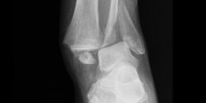
Osteochondral lesions of the talar dome
The incidence of such lesions have been reported to be up to 70% in cases of acute ankle injury especially ankle fractures. Patients usually present with persistent pain, Early on swelling and bruising with limitation in the motion available in the joint. Later on a dull ache and stiffness may be the only signs. Clicking and locking can also occur. If conservative measures fail to improve the symptoms and disability operative treatment may be recommended in the form of excision, curettage and drilling of the lesions if amenable to it.
Arthroscopy can be used as a diagnostic modality but also for the treatment of:
-
Loose bodies
-
Soft tissue impingement
-
Synovial Disorders: RA, PVNS, synovial chondromatosis, Hemophilia.
-
Osteochondral lesions of the talar dome
-
Tibiotalar osteophytes; bony impingement
-
Arthroscopic arthrodesis for arthritis of the ankle
-
Arthroscopic repair for ankle instability
-
Os trigonum or posterior hindfoot impingement